Elemental calcium 2-4g per day +\- rocalcitrol 0.25 ug qd
Calcium carbonate: (1g CaCarbonate has 400mg elemental calcium)
If symptomatic 1 amp calcium gluconate in 50ml d5w over 20 min
Replace magnesium
Monday, January 11, 2010

Tuesday, January 5, 2010
Exposure Abdominal Aortic Aneurysm
1) Draping: shave abdo and groins, knees to nipple prep, groin drape and square offs, ioban over abdomen
2) Midline incision from xiphi to pubis
3) Check placement of NG
4) Find and divide LT and mobilize 4th part of duodenum by dividing filmy attachment between duodenum and inferior mesenteric vein
5) Place bookwalter or omni with right sided bias as most contents will be retracted that way
6) Retract tranverse colon by placing splancnic retractors to also retract duodenum and IMV
7) Place large maleable retractor at base of small bowel mesentery and retract to right
8) Place maleable retractor in RLQ and LLQ
9) small side wall retractor for abdominal wall to left
Glasgow Coma Scale (GCS)
Adults: EVM (456):
Eyes:
1- none
2- open to pain
3- open to voice
4- spontaneously
Verbal:
1- none/intubated
2- incomprehensible
3- inappropriate
4- confused
5- oriented
Movement:
1- none
2- decorticate posturing
3- decrebrate posturing
4- withdraws from pain
5- localizes to pain
6- spontaneous
Eyes:
1- none
2- open to pain
3- open to voice
4- spontaneously
Verbal:
1- none/intubated
2- incomprehensible
3- inappropriate
4- confused
5- oriented
Movement:
1- none
2- decorticate posturing
3- decrebrate posturing
4- withdraws from pain
5- localizes to pain
6- spontaneous
Monday, January 4, 2010
Buerger's Disease
AKA: Thromboangitis obliterans
- non-atherosclerotic, segmental inflammatory disease that affects small and medium sized arteries and veins of the extremities.
- affected patients are almost uniformly smokers to the point where somking is more or less a pre-requisite for this diagnosis.
Presentation:
- ischemia of distal small arteries and veins
- claudication of distal extremities, two or more limbs always involved
- differentiate from claudicants who have calf cramping typically
- progression leads to ischemic rest pain and ulceration of fingers and toes
Diagnosis:
- rule out: scleroderma, CREST, mixed connective-tissue disease, SLE, hypercoaguability disorders with serum screens (CBCD, lytes, BUN, Cr, LFTs, BG, UA, sed rate, CRP, hypercoagubility screen incl APAb, ANA, RF, Complement, SCL-70, anticentromere Ab)
- angiography (MRA or CTA) may be needed
- echo to rule out proximal embolic source
Treatment:
- stop smoking!!
- limited options if arterial occlusion has developed +/- ulceration
- calcium channel blockers may be useful if pt has developed Raynaud's phenomenon
- Revascularization is not an option as distal targets and saphenous conduits are toast
- Although graft patency rates are low if targets are available, if it heals ulcer usually results in high rate of limb salvage.
- Amputations inevitable in pts with extensive gangrene or sepsis
Lymphedema
Occurs when there is impaired uptake of lmphatics
- increases interstitial oncotic pressures and perpetuates egress of lymph fluid
- remaining lymphatics dilate and develop valvular incompetence over time
- fibrosis of lymphatic walls eventually obilterates lymphatic channels
Skin thickens over time if lymphedema is not treated
- epidermis develops thick scaly deposits
- cracks in thickened epidermis can lead to cellulitis/lymphangitis
- may progress to malignant degeneration to lymphoscarcoma (rare): Stewart-Treves syndrome
- Lymphoscarcoma presents with reddish-purple discoloration or nodule, tends to form satellite lesions
- treatment of lymphosarcoma involves radical amputation, poor prognosis
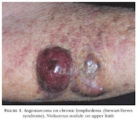
Stewart-Treves Syndrome
Etiology:
- primary vs. secondary
Primary:
- all types more common in females
- Lymphedema I (Millroy's disease): present at birth, affects dorsum of foot, not typically progressive
- Lymphedema II (Lymphadema Praecox/Meige's disease): onset at puberty, most common form of lymphedema (65-80%). Affects lower extremities, unilateral. Peripheral lymphatics are hypoplastic.
- Lymphedema III (lymphedema tarda): presents in midlife (>35yo). Lower extremities.
Secondary:
- infection, cancer, morbid obesity
- worldwide, most common cause is infection with parasite (Wuchereria bancrofti)
Diagnosis:
- basic history and physical
- rule out other causes of lymphedema (cardiac, venous, renal, hepatic, iliac compression)
- if imaging desired then lymphoscintigraphy can be used to demonstrate decrerased clearance of lymph
Treatment:
conservative management predominates
- stop the positive feedback cycle of inflammation, infection and fibrosis
- skin hygeine, treatment of infection
- massage protocols to decompress limb followed by compression stockings if arterial supply is sufficient
medical treatment is limited:
- Abx to treat infection
- use of diuretics and benzopyrene (increase proteolysis in edema) are of questionable benefit
surgical management is described but rarely of benefit:
- debulking procedures (reduce size and weight of limb) - however, will also disrupt what lymph channels remain
- physiologic procedures attempt to reestablish lymphatic flow - long-term outcomes not good overall.
- increases interstitial oncotic pressures and perpetuates egress of lymph fluid
- remaining lymphatics dilate and develop valvular incompetence over time
- fibrosis of lymphatic walls eventually obilterates lymphatic channels
Skin thickens over time if lymphedema is not treated
- epidermis develops thick scaly deposits
- cracks in thickened epidermis can lead to cellulitis/lymphangitis
- may progress to malignant degeneration to lymphoscarcoma (rare): Stewart-Treves syndrome
- Lymphoscarcoma presents with reddish-purple discoloration or nodule, tends to form satellite lesions
- treatment of lymphosarcoma involves radical amputation, poor prognosis
Stewart-Treves Syndrome
Etiology:
- primary vs. secondary
Primary:
- all types more common in females
- Lymphedema I (Millroy's disease): present at birth, affects dorsum of foot, not typically progressive
- Lymphedema II (Lymphadema Praecox/Meige's disease): onset at puberty, most common form of lymphedema (65-80%). Affects lower extremities, unilateral. Peripheral lymphatics are hypoplastic.
- Lymphedema III (lymphedema tarda): presents in midlife (>35yo). Lower extremities.
Secondary:
- infection, cancer, morbid obesity
- worldwide, most common cause is infection with parasite (Wuchereria bancrofti)
Diagnosis:
- basic history and physical
- rule out other causes of lymphedema (cardiac, venous, renal, hepatic, iliac compression)
- if imaging desired then lymphoscintigraphy can be used to demonstrate decrerased clearance of lymph
Treatment:
conservative management predominates
- stop the positive feedback cycle of inflammation, infection and fibrosis
- skin hygeine, treatment of infection
- massage protocols to decompress limb followed by compression stockings if arterial supply is sufficient
medical treatment is limited:
- Abx to treat infection
- use of diuretics and benzopyrene (increase proteolysis in edema) are of questionable benefit
surgical management is described but rarely of benefit:
- debulking procedures (reduce size and weight of limb) - however, will also disrupt what lymph channels remain
- physiologic procedures attempt to reestablish lymphatic flow - long-term outcomes not good overall.
Short Saphenous Vein
- passes behind distal end of fibula (lateral malleolus) up the back of the leg to penetrate the deep fascia and join the popliteal vein
- medial sural cutaneous closely approximates the short saphenous and care must be taken to avoid it (Plate 468 Netter's)
- medial sural cutaneous nerve harvested as a cable graft for facial nerve when resected along with parotid.

- medial sural cutaneous closely approximates the short saphenous and care must be taken to avoid it (Plate 468 Netter's)
- medial sural cutaneous nerve harvested as a cable graft for facial nerve when resected along with parotid.
Nutrition Academic Half-Day:
Dumping Syndrome:
- 4-6 small meals a day
- Avoid liquids with meals
- liquids 30 mins after a meal
- avoid high sugar foods
Bariatrics:
- full fluids for 2 weeks post-op
- avoid carbonated drinks, caffeine, EToH
- high protein choices
- 1/2 cup fluids while pt awake
- transition to solids 2 weeks post-op
- 3 meals with snacks in between
- drink milk between meals to incrase Ca and protein intake
- start 1/4 cup at a time, in crease to 1 cup (6-8 weeks)
- no drinking with meal/solids (so they don't fill up with low nutrient fluids)
- daily multivitamin
- calcium citrate with Vit D (Citrate is only form of calcium absorbed in the stomach)
- 1200-1500 IU Ca
- 800 U Vit D
Subscribe to:
Posts (Atom)